 RSS Subscribe
Welcome to the new world of regenerative medicine: the plug and play human body – Richard Mills
2026.07.30
One of the most perplexing issues to confront modern medicine has been the successful transplantation of human organs.
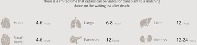
The problem isn’t so much the procedure — although there is always the risk of the recipient’s body rejecting the organ, hence the use of immunosuppressive drugs — but the fact that demand vastly outstrips supply. Transplant patients get placed on a waiting list for an organ, making the success of the system entirely dependent on the number of organ donors. The donor has to have the right blood type and the organ must be the right size for there to be a match. Many patients become sicker as they wait; in every country, hundreds die each year waiting for an organ.
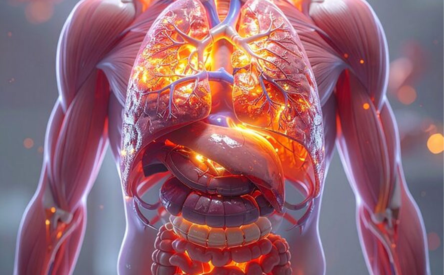
Organs that can now be transplanted include the liver, kidney, pancreas, heart, lung, intestine, cornea, middle ear, skin, bone, bone marrow, heart valves and connective tissue.
According to the United Network for Organ Sharing, every day 21 people in the United States die waiting for an organ, and over 120,000 people are on organ transplant waiting lists.
According to the Canadian Institute for Health Information (CIHI), almost a third (31%) of 678 Canadians who in 2025 were removed from organ transplant wait-lists, had died while waiting.
While Spain has 43 donors per million people, the US has 26, Britain has 21, and Canada has just 20. A few more facts about organ transplants in Canada:
- Only 1% of deaths in hospitals yield organs for transplants
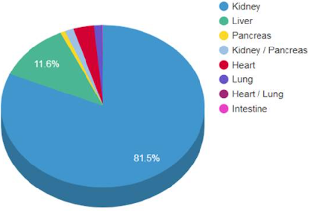
- 75% of the waiting list are kidney recipients. The next in-demand organs are the liver, lungs and heart
- Ontario is estimated to spend $100 million a year caring for sick patients awaiting a kidney transplant
- The average kidney transplant patient will wait four years
- Over 1,600 people are added to waiting lists each year
Imagine if, instead of waiting for an organ from another person —possibly a relative but likely a stranger — you could walk into a clinic and have one manufactured with your cells. It sounds far-fetched, but the technology now exists for the tailor-made transplantation of organs through brand-new medicine called 3D bioprinting.
What is 3D bioprinting?
Most people are familiar with the relatively new field of 3D printing. Put simply, 3D printing is a progression of 2D printing, where a third dimension is added to the printing of images on a flat surface (a regular ink-jet printer), adding depth and allowing the printer cartridge to move in all directions. A digital file is first created using modeling software, then sent to the printer, depositing layers of the chosen material — often plastic or wax — to build up the final product.
Formerly known as stereolithography, 3D printing was invented in 1983 by Chuck Hull, co-founder of 3D Systems. It took over 30 years for the technology to become mainstream, but now 3D printing can be done by anyone with access to a 3D printer, which can be purchased for under $500.
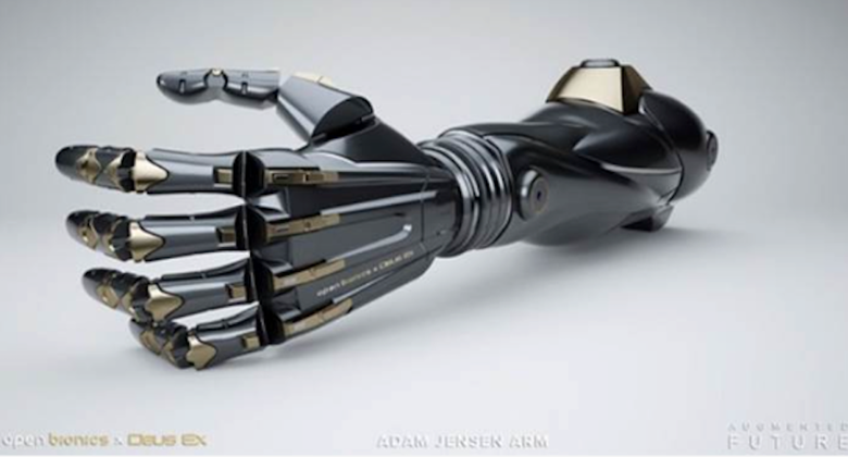
Among the more interesting items that have been 3D-printed are prosthetic limbs, fabricated firearms, electrical vehicles, steel parts (Caterpillar introduced the first 3D-printed excavator in 2017), quick-build homes, parts for combat aircraft and spacecraft, and even plant-based meat alternatives.

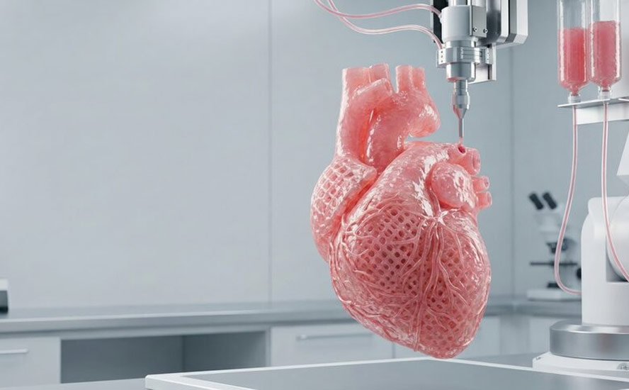
Bioprinting operates on the same general principle as regular 3D printing but instead of plastic, wax or other matter, bioprinters deposit layers of living cells to build structures like blood vessels or skin tissue. The cells are taken from an animal or a human being and cultivated until there are enough to create “bioink” which is then loaded into the printer using mechanical syringes. Adult stem cells can also be utilized.
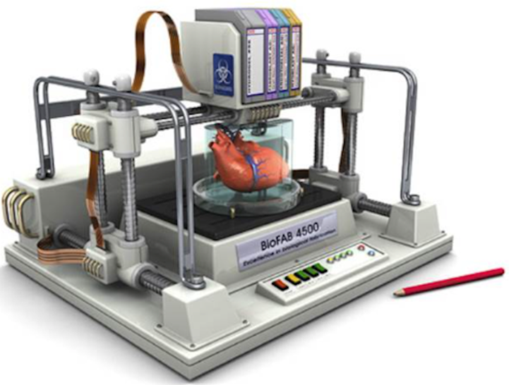
Key to the process is a dissolvable gel which acts as a kind of incubator for the cells to multiply — like an embryo growing in a womb. Researchers may also plant cells around 3D scaffolds made of biodegradable polymers or collagen, allowing them to develop into functional tissue. The cells use their inherent properties to seek out similar cells to join with. Researchers are able to control the shape into which the cells form, and the printer builds the final structure.
After the tissues are fully grown and shaped, they are placed into a recipient’s body. The hope is that the 3D-printed object becomes as much a part of the patient’s body as the cells he or she was born with.
Dr. Utkan Demirci, professor at Stanford University School of Medicine, defines bioprinting as the process of using advanced additive manufacturing technologies to pattern biological materials, such as cells, biomaterials and biomolecules, for the fabrication of tissue-mimicking constructs. This novel approach requires biocompatible materials called bio-inks to act as the matrices for printed cells, which can then be grown in bioreactors to further develop and become functionally mature.
— PhysicsWorld: The past, present and future of 3D bioprinting
There are currently five common methods of 3D bioprinting:
Inkjet bioprinting: Droplets of bio-ink are deposited, layer by layer, onto a culture plate. Cells that can help fight breast cancer have been successful printed using inkjet bioprinting.
Extrusion bioprinting: Polymer or hydrogel is loaded in syringes and dispensed via pneumatic- or screw-driven force, onto a building platform. The motion is controlled by a computer. Extrusion bioprinting offers lower resolution than inkjet bioprinting but the fabrication speed is considerably higher, allowing anatomically-shaped objects to be generated.
Laser-assisted bioprinting: A laser is used to deposit the biomaterials into a receptor via a tape covered with biological material. The laser irradiates the tape, causing the biological material to evaporate and reach the receptor in the form of droplets. The droplets contain a biopolymer that acts as an adhesive to help the cells to grow. This high-resolution bioprinting method is being used in a partnership between French bioprinting company Poietis and L’Oréal to recreate a hair follicle that could lead to a cure for baldness.
Stereolithography: Stereolithographic bioprinting uses a “digital micromirror” to direct ultraviolet light onto the printing surface. Light directed by the micromirrors triggers the formation of molecular bonds, which cause light-sensitive hydrogels to form into solid material.
Bioprinting with acoustic waves: Using a device that allows cells to be manipulated with acoustic waves, researchers can manipulate where the waves will meet along three axes. The waves then form a trap that captures the cells, which are collected to create 3D patterns.
How far has it progressed?
Bioprinting has advanced from printing simple cell layers to creating functional, complex 3D structures like skin, bone, and blood vessel networks, though fully functional printed organs for routine transplants remain in development.
Scientists routinely print skin, cartilage, and bone tissue used in grafts and orthopedic care.
Techniques now allow printing of tiny blood vessel channels needed to keep thick, complex tissues alive.
New methods using cell clusters (spheroids) build tissue patterns up to 10 times faster than older techniques.
Lab-grown human tissue models test new medicines safely without using animals.
Simple patches for hearts and engineered corneal tissues are moving closer to or entering human testing phases.
Emerging tech aims to print skin or bone directly onto a patient’s wound during surgery.
Some of the most advanced work on bioprinting has been done at the Wake Forest Institute for Regenerative Medicine in California. One of the first major structures that Wake Forest bioprinted was a human bladder. Made from cells extracted from a patient with a poor-functioning bladder, the 3D-printed bladder was successfully transplanted. The project built on custom-grown bladders that had previously been transplanted into seven patients suffering from spina bifida, a birth defect that affects the spinal cord.
Wake Forest staffers have also created an outer human ear, and implanted bioprinted skin, bone and muscle on laboratory animals that successfully grew into surrounding tissue.
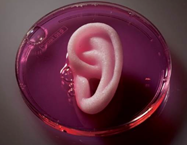
But these are just the early prototypes. Bioprinting’s leading researchers expect that as the field develops, the technology will evolve into the ability to bioprint major organs for transplant into human donors — completely eliminating waiting lists, patient deaths, and dramatically increasing the success rate of organ transplants.
The institute’s director, Anthony Atala, sees bioprinting as totally transforming the relationship between the transplant patient and doctor, in much the same way that Dell changed the way consumers interacted with the computer company that sold PCs tailored to each customer’s unique needs. Patients could order replacement parts in much the same way they might order a new clutch for their Mazda.
“You’d have companies that exist to process cells, create constructs, tissue. Your surgeon might take a CT scan and a tissue sample and ship it to that company,” Atala said in a feature article on bioprinting in Smithsonian Magazine.
The company would then ship the organ back a week or so later, ready for implantation. Welcome to the new world of regenerative medicine: the plug and play human body.
Atala said the technology is developing to the point where researchers are almost able to replicate simple organs like the outer ear and the trachea (windpipe). Importantly, there are no real surgical challenges, he told Smithsonian.
Skin
Wake Forest is working on a skin-cell printer capable of printing live skin cells directly onto a burn wound. The procedure could replace skin-grafting, a procedure where healthy skin is harvested from an unburnt part of a patient’s body. Skin grafting can be hard to heal from, and in severe burn cases, there isn’t enough healthy skin left to use.
This new printing technique only needs a patch of skin 10% the size of the burn, that is used to grow enough cells for 3D printing. The wound is then scanned for size and depth, information which the printer uses to print skin cells at the proper depths to cover the wound.
Recent developments in 3D bioprinting skin focus on robotic in-situ wound printing, multi-layered humanized tissue models, and advanced bioinks. These innovations aim to replace traditional painful skin grafts by directly reconstructing full-thickness skin layers, blood vessels and appendages.
Hearts
At the Texas Heart Institute in Houston, researchers are working with decelluarized pig hearts. The organs have been stripped of muscle and other living tissue, but the original architecture is intact. The idea is to use decelluarized pig hearts, repopulated with bioprinted human cells, for implantation into humans. So far the institute has succeeded in injecting pig hearts with living bovine cells, then inserted them into cows where they worked successfully next to a cow’s heart.
Already, patients with a defective heart valve can have a pig’s valve or a mechanical valve implanted. Doris Taylor, director of the institute’s regenerative medicine research program, says the decelluarized method gets around the tricky process of printing at the extremely high resolution required for highly vascularized (containing many blood vessels) organs like the heart.
“The tech is going to have to improve a great deal before we’re able to bioprint a kidney or a heart, and get blood to it, and keep it alive,” Taylor told Smithsonian.
Developments though are moving in that direction. In 2016 Harvard researchers 3D-printed the first “heart-on-a-chip”. The tiny device contains living human heart cells that mimic the heart’s functions.
In 2018, 3D printing startup BioLife4D successfully produced human tissue in the form of a cardiac patch — derived from a patient’s white blood cells with multiple cell types contained in the human heart. According to pharmaforum, it’s another step towards bioprinting major organs for transplant.
Scientists at the American Friends of Tel Aviv University have reportedly 3D-printed a fully-vascularized heart using fat cells from a donor. The fat cells were partially cultured and re-programmed into heart cells. This early-stage technology has only been able to print a heart the size of a rabbit’s, but researchers hope to test the printed hearts in other animals.
Recent breakthroughs in bioprinting hearts focus on printing with clusters of stem cells called organoids, 4D shape-morphing architectures, and embedded vascular networks. These innovations improve cell alignment, tissue contraction and nutrient delivery, bringing scientists closer to functional cardiac patches.
Ovaries
Northwestern University in Illinois debuted a 3D-printed ovary using the acoustic waves method described above, and in Sweden, researchers have successfully created human cartilage tissue, also using acoustic waves.
Recent breakthroughs in ovarian bioprinting focus on advanced bioinks and architectural scaffolds, key developments that improve hormone restoration and follicle survival. These innovations aim to help cancer survivors and patients with premature ovarian failure regain reproductive and endocrine health.
Thyroids
Russian scientists aboard the International Space Station successful bioprinted the first organ in space: a mouse’s thyroid. Space’s zero-gravity environment enables tissues to mature faster than on Earth.
Recent developments in thyroid bioprinting focus on microfluidic printing technologies, decellularized extracellular matrix hydrogels, and high-throughput endocrine testing systems. These innovations improve how scientists model the thyroid gland and test for chemical safety.
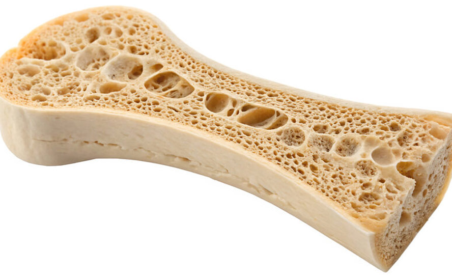
Bones/ cartilage
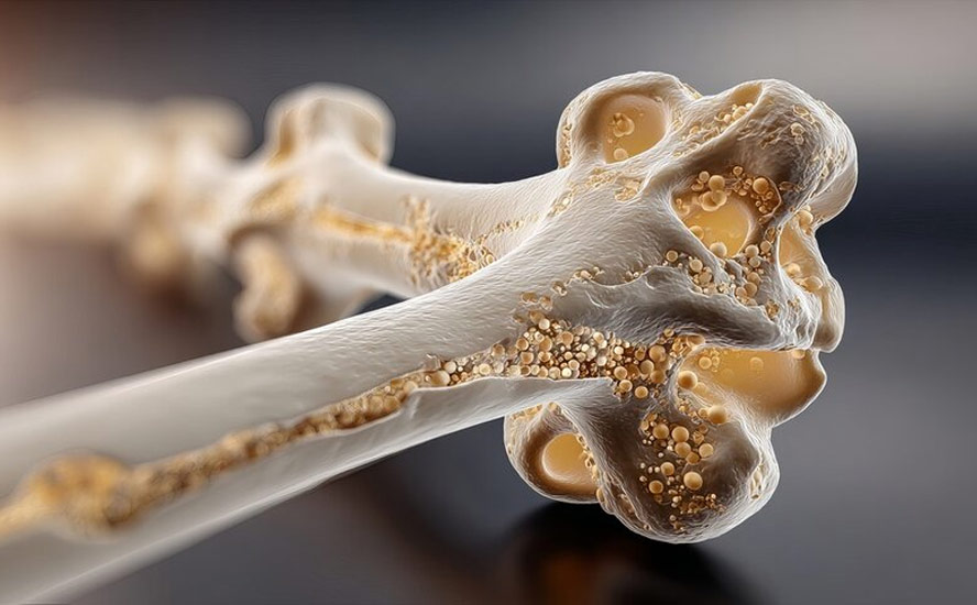
Supported in part by the National Science Foundation, researchers at Penn State University are working with a team that is bioprinting bone, encoding two growth factor genes to enhance healing and to regenerate bone. Bioprinting bones could one day accelerate the healing process of broken bones.
Meanwhile, a team of scientists in Gothenburg, Sweden are hoping to use bioprinted cartilage in patients with muscle issues. The first patients would likely be those with sports injuries. The technology could also be applied to elderly patients, such as those who suffer from osteoarthritis.
Also, a team from the UK’s Swansea University has developed a bioprinting process that uses regenerative material to create an artificial bone matrix. The technology could replace bone grafting, a surgical procedure that replaces missing or damaged bones with synthetic materials. Unlike bone grafting, which doesn’t allow new bone tissues to form, thus limiting mechanical integrity, 3D-printed bones are capable of fusing with, and even replacing over time, a patient’s natural bones.
Cartilage printing could revolutionize joint care through a hand-held cartilage printing device called BioPen. Built by Australian researchers, the BioPen contains stem cells derived from a patient’s fat, which create “custom scaffolds of living material into failing joints” much like 3D-printed bones.
The device has successfully generated human cartilage in laboratory studies and regrown cartilage in sheep, though it has not yet completed human clinical trials.
Corneas
Finally, a group of researchers in South Korea has 3D-printed prototype corneas from decelluarized corneal stroma and stem cells. Unlike artificial corneas currently available, made of substances like synthetic polymer which resist incorporation into the eye, printed corneas are made to mimic the material within natural corneas. The invention could replace the need for donors and synthetic corneas in cataract surgery and other sight complications.
Recent developments in corneal bioprinting feature ultra-fast printing times under 10 minutes, advanced decellularized extracellular matrix (dECM) bioinks, and patient-specific digital eye-scan matching. These innovations aim to bypass global donor tissue shortages.
AI and 3D bioprinting
Artificial intelligence improves 3D bioprinting by optimizing bioink formulations, converting patient scans into precise tissue models, and providing real-time quality control during printing.
AI turns patient CT or MRI scans into custom 3D digital blueprints for implants.
Machine learning designs complex internal pathways, like tiny blood vessels, that mimic real body tissues.
AI predicts how mixtures of living cells and gels will flow, hold their shape, and support cell growth.
Cameras and sensors watch the printing process live, automatically fixing tiny errors or nozzle clogs in milliseconds.
Leading companies using artificial intelligence for 3D bioprinting include Aspect Biosystems, Rokit Healthcare and BioLattice. These pioneers use machine learning and computational design to program cells, optimize bioinks and automate tissue manufacturing.
A Belfast start-up that produces 3D prints of body parts is developing a machine-learning technique to automate the process of taking 2D images of slices of the anatomy to build up a 3D model. The images are necessary to denote bone, muscle and organ tissue. Automation would replace this task, currently performed by medical visualization engineers, which can take up to four hours per print. Because there are common features to human anatomy, a machine could be trained to scan previous files and label images.
Quantum computing and 3D bioprinting
Quantum computing is an advanced technology that uses the rules of quantum physics to solve complex problems much faster than normal computers.
Quantum computing is used in 3D bioprinting mainly to optimize bioink molecular formulas, simulate complex cellular microenvironments, and calculate intricate multi-nozzle printing pathways. While the physical printing hardware relies on classical mechanics, quantum algorithms help solve intensive biochemical variables that bog down regular computers.
Quantum computing models complex atomic interactions in hydrogels and biomaterials at an unprecedented scale.
It predicts how adjustments to bio-inks will affect cell survival, viscosity, and tissue development before physical lab testing.
And it rapidly scans vast combinations of biomaterials to find optimal tissue-scaffolding recipes.
Quantum algorithms process multiple nozzle path configurations simultaneously to prevent structural collapse in soft constructs.
They reduce placement errors during the deposition of delicate living cells, improving the overall reproducibility of complex tissue layers.
What are the challenges?
Printing large, multi-layered solid organs like a functioning human heart or kidney remains a major long-term challenge.
Keeping thick, engineered tissues alive and fully integrated with a patient’s natural blood supply over time requires more study.
Clear global safety rules and standardized manufacturing guidelines are still being built.
Immunity is a major challenge in 3D bioprinting, primarily involving host rejection of biomaterials, foreign body reactions, and the difficulty of accurately replicating complex immune microenvironments.
The hydrogels and polymers (bioinks) used to structure printed tissues can trigger inflammation, immune cell infiltration, and fibrous encapsulation by the host.
Implanted bioprinted constructs face adaptive and innate immune responses similar to traditional organ transplants, risking rejection unless patient-specific cells or immunomodulatory designs are used.
Engineering functional 3D models that successfully simulate active immune responses, such as macrophage polarization or lymphocyte infiltration, remains difficult due to current bioink and structural limitations.
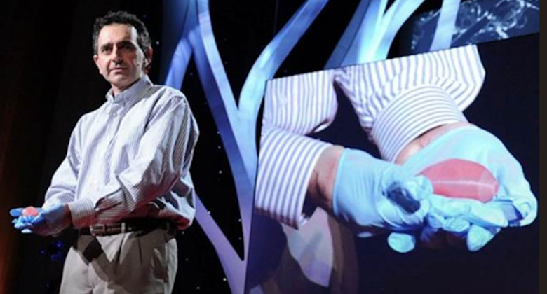
The holy grail of 3D bioprinting would be to come up with a viable kidney for transplant. Prof. Atala, of the Wake Forest Institute, created the first small-scale bioprinted kidney in 2002. However, Atala is the first to admit that his machine-produced kidney is nowhere near at the level it needs to be for a human transplant. A TED Talk Atala gave in 2011 about bioprinting, which culminated with a dramatic display of an object — really an over-sized bean — became controversial when the press got ahold of it and printed enthusiastic, but wrong, stories about the technology eliminating the need for a kidney transplant.
Another potential roadblock is the cost. No-one yet knows what it would cost to bioprint and transplant a human organ on demand, and how accessible the procedure would be to the masses of patients requiring a transplant. And while there have been successful bioprinted organ transplants, there haven’t been enough to determine how well the human body will accept the new tissue or artificial organ.
Finally, one shouldn’t underestimate the complexity and level of difficulty involved.
Describing the complexity of a human kidney, The Smithsonian Magazine article indicates why bioprinting still has a ways to go:
Sit down with a pencil and a piece of paper and you could hardly dream up something more architecturally or functionally complex than the human kidney. The interior of the fist-size organ is made up of solid tissues traversed by an intricate highway system of blood vessels, which measure as little as 0.010 millimeters in diameter, and approximately a million tiny filters known as nephrons, which send healthful fluids back into the bloodstream and waste down to the bladder in the form of urine. To bioprint a kidney, you’d have to be able to cultivate and introduce not only functioning kidney cells and nephrons, you’d also need to have mastered how to populate the organ with a vasculature to keep the organ fed with the blood and nutrients it needs. And you’d have to build it all from the inside out.
As pharmaforum points out, “A complex network of cells, tissues, nerves and structures in a human organ need to be correctly positioned with a highest precision for it to function properly. From arranging the thousands of tiny capillaries in a liver, to printing a heart that beats, it is a long, difficult process.
While some parts of the human body are more complex than others, each piece has its own specialised requirements and issues that need addressing. The selection of the right materials, cell types and bio-inks must be as precise as the blueprint itself.”
Complete, functional 3D-printed internal organs are still 15 to 30 years away from regular clinical use, though simpler structures like skin, cartilage, and ears are already entering early clinical trials, states CELLINK, a leading bioprinting company.
Indeed, patients may need to be satisfied with machine-engineered tissue rather than organs, according to The Medical Futurist:
Synthetic skin, a bionic ear, bladder, or cornea might be the first tissues to be either bioprinted or grown in the lab on demand – since they are tissues containing a small number of cell types. After that, more complicated ones might be engineered.
Investment opportunity
Still, 3D bioprinting has come a long way since Atala’s first artificial bladder in 2002. At Ahead of the Herd, we think it is the next big thing in regenerative medicine. Science always starts out with experimentation, sometimes many years of it, before the technologies are commercialized. We want our subscribers to be well aware of 3D bioprinting’s potential, putting them in a position to get in early to companies that are offering bioprinted products.
We see an entire ecosystem of small firms developing, with each focusing on a different aspect, technology or part of the body. It will not take 10 years for start-up public companies to IPO, seeking money to develop their technologies.
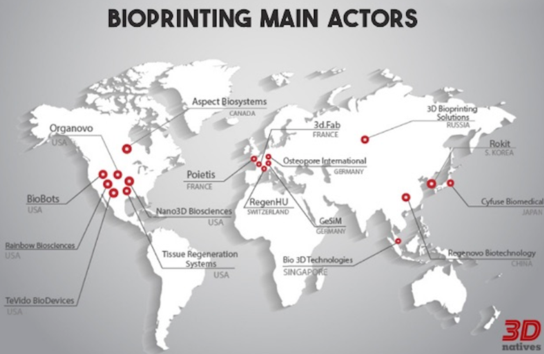
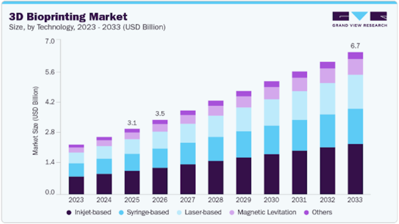
The global 3D bioprinting market size was valued at approximately USD$3.1 billion in 2025 and is projected to reach $3.5 billion in 2026, heading toward roughly $6.7 billion by 2033. Industry trackers estimate the compound annual growth rate (CAGR) will run between 9.7% and 12.7% over the coming decade.
North America holds the largest revenue share (28-38% of the market) due to high biomedical R&D spending. This makes 3D bioprinting an ideal new sector for Canada and US-focused investors.
Conclusion
3D bioprinting could represent the solution to the transplant dilemma that has plagued medicine for hundreds of years. People are dying waiting for organs, or in severe pain. Imagine if that pain could go away, literally, in a heartbeat. While it’s still early days as far as getting to the transplantation of major organs, the potential for this technology is enormous. Instead of patients being hardwired into an organ transplant system that is slow, risky and heartbreaking for those who are too far down the waiting list, 3D bioprinting holds the promise of medicine tailored to the individual. We are on the cusp of the human body being plug and play: get a scan of your defective organ, and a tissue sample, then send it off to a company for organ fabrication. A week later, presto! New organ, problem solved.
3D bioprinting and quantum computing are high on my radar as I hunt for new AOTH investment opportunities.
Richard (Rick) Mills
aheadoftheherd.com

Legal Notice / Disclaimer
Ahead of the Herd newsletter, aheadoftheherd.com, hereafter known as AOTH.Please read the entire Disclaimer carefully before you use this website or read the newsletter. If you do not agree to all the AOTH/Richard Mills Disclaimer, do not access/read this website/newsletter/article, or any of its pages. By reading/using this AOTH/Richard Mills website/newsletter/article, and whether you actually read this Disclaimer, you are deemed to have accepted it.